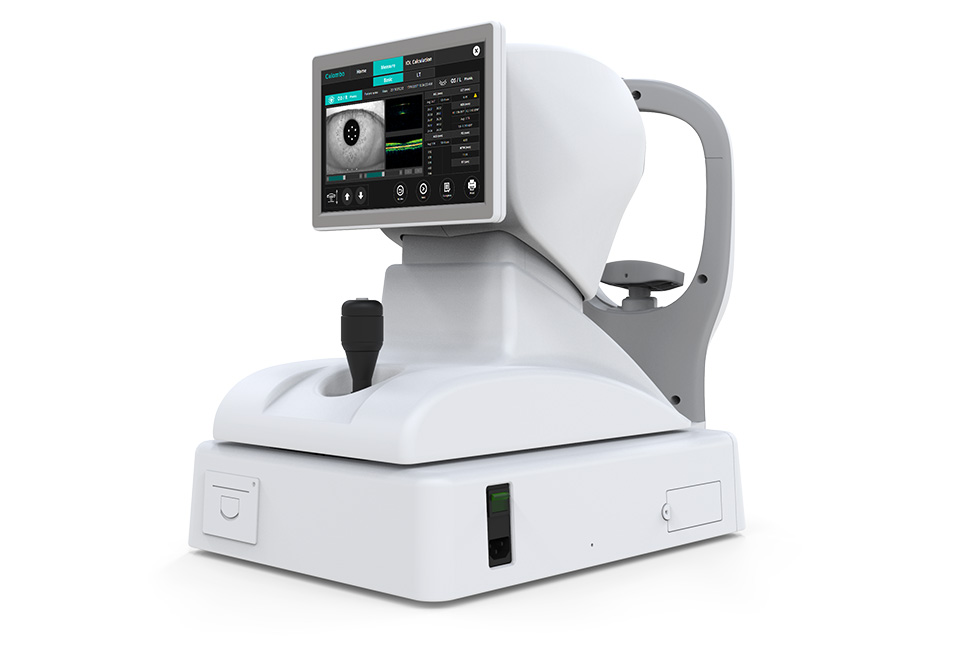
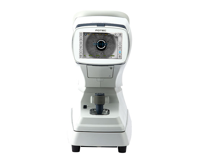
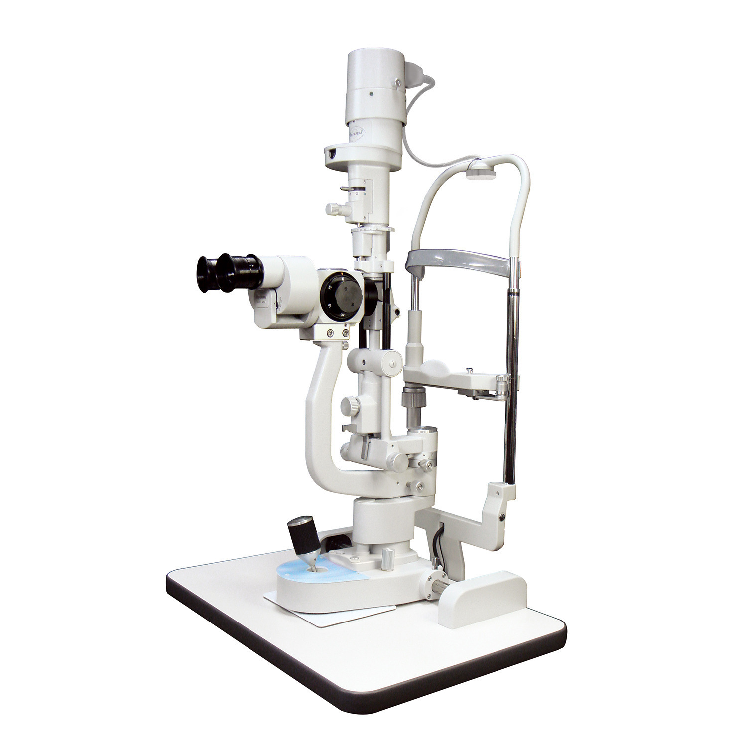
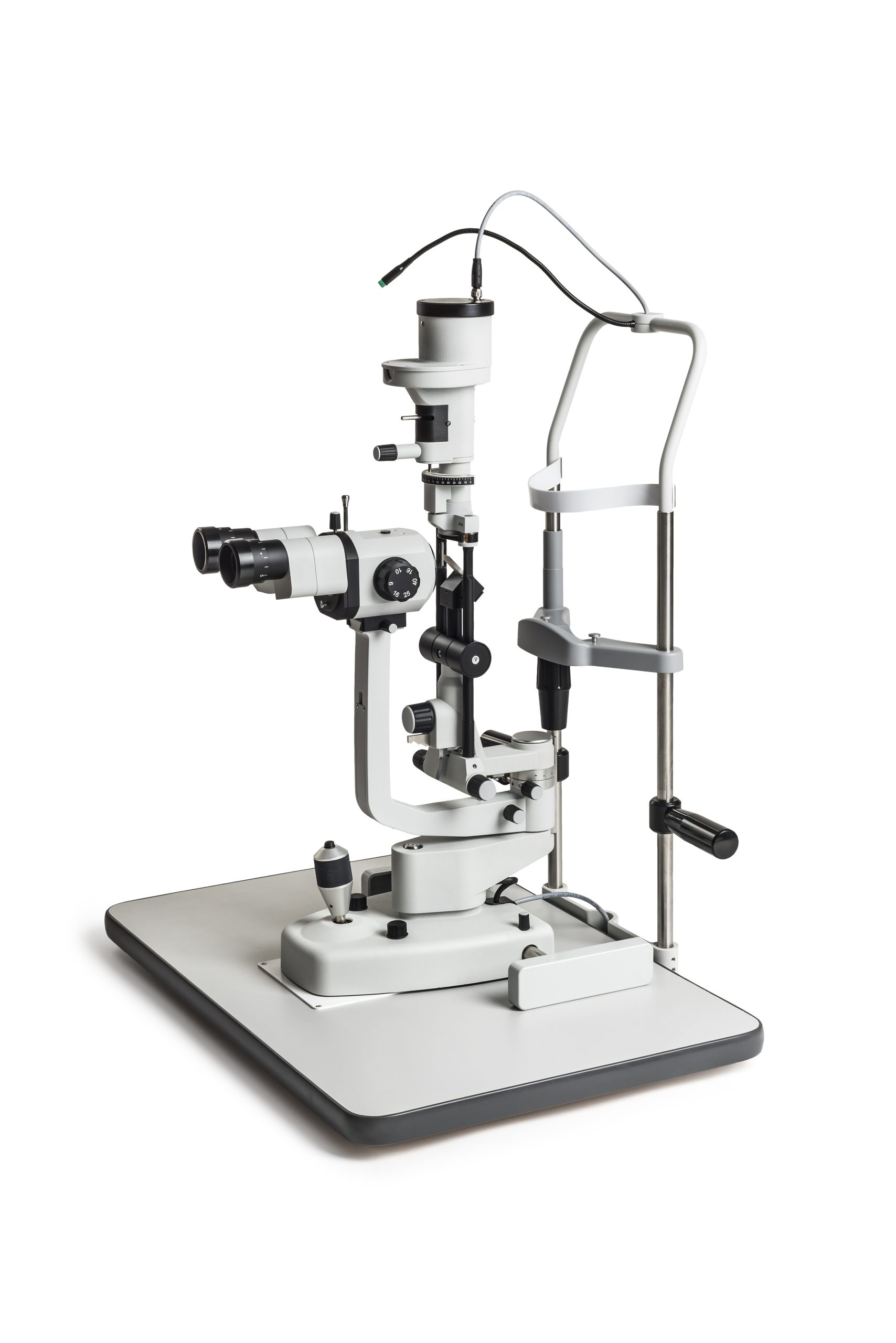
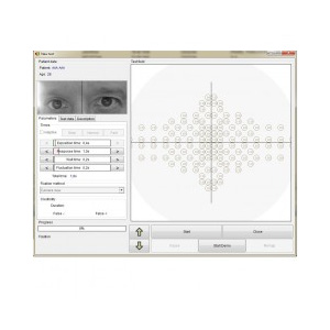
VISUAL fIELD ANALYZER AP-50

Category:

AP-50 is a desktop model, lightweight and fully featured modern static automated perimeter ideal for glaucoma diagnosis and specific requirements of occupational medicine and busy mobile clinicians. AP-50 uses LED back projection of stimulus in white color, and offers a wide range of strategies, test fields and reach set of test parameters to assure quick and precise measurement. Control of fixation is performed automatically using the built-in camera or by controlling the position of the blind spot. Built-in data analysis include regression analysis and standardized ways of presenting and printing examination results. Perimeter AP-50 can work with any PC computer running the Windows operating system.

Reduced device size thanks to novel measurement bowl design

Elliptically shaped measurement bowl realizes significantly reduces size of thedevice yet diagnostic value of AP-50 is comparable to full field perimeters.Full central 30 degree of patient vision with extension to 50 degree temporallyallows precise test nasal step where early glaucoma often commences early in the disease progression.FREY Visual Field Analyzer - AP-50

Small size, compact and light weight

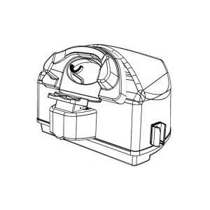
AP-50 dimensions of only 548 x 382 x 450 mm and weight only 9kgmakes the device an ideal desk top clinical tool which can be easilytransported, set up and installed anytime, anywhere including remote sites.

FREY Visual Field Analyzer - AP-50

Less demanding for ambient light conditions

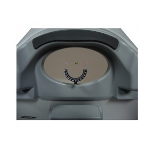
Elliptically shaped measurement bowl requires lower test roomillumination conditions. Electronically controlled backgroundillumination assures stable measurement conditions.FREY Visual Field Analyzer - AP-50

Standard test fields and conditions

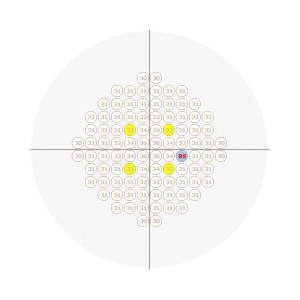
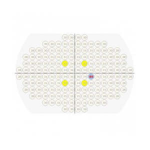
Standard test fields like Central 30, Central 24, Macula are availableall allow for easy data comparison with other perimeter systems.Precise testing in nasal step dedicated glaucoma tests are included.FREY Visual Field Analyzer - AP-50Binocular testing
AP-50 fulfils specific visual field testing requirements for driversand occupational medicine. Measurement range covers up to 160 degrees temporally,and both eyes can be tested simultaneously

AP-50 TECHNICAL SPECIFICATION
Device type
Automated perimeter
Measurement bowl type
Hemispherical 300mm radius with difusive surface
Maximum temporal range (degrees)
80°
Stimulus
White-on-white
Stimulus duration
0.1 – 9.9s
Stimulus size
III
Stimulus intensity
0.03asb to 10000asb
Visual field testing distance
30cm
Background illumination

White 31.5asb (10cd/m2)

Automatic background illumination control

Fixation control

Heijl-Krakau blind spot monitor

Eye tracking (video camera)

Eye previev (video camera)

Test models

Supra threshold age corrected: screening

Threshold: Full Threshold, Fast Threshold, Smart Threshold
Others: 2-Zone, 3-Zone, Quantify Defect, Constant, Binocular, Bi-Driving, Targeted Perimetry, Neurological

Test field library

Macula, Central 10°, Central 20°, Central 30°

Nasal step (Glaucoma)

Correction glass diameter
38mm
Chinrest
Electrically driven in horizontal and vertical axis
Computer
Touch screen support
Printer
External or network printer
Additional software features

Fovea threshold testing

Automatic pupil measurment

User management module

Touch screen operation

DICOM export

Network connectivity

Programming interface for EMR systems

Data import from HFA devices

Auto backup

Dimensions

Height: 382 mm
Width: 548 mm
Depth: 450 mm

Weight
9kg
Power requirements

Voltage 110-230VAC

Power 65W

Intuitive user interface

User interface of Perimetry application is designed to suport efficient work with minimum time spent on switching between options. It’s ergonomy and simplicity assures short learning time for the operator. The application is ready to be operated with use of touch screen or with mouse and keyboard depending on user preferences.

Defect progress analysis

Regression analysis module allows user to track changes in field of vision in time, using easy for interpenetration graphs. Multiple examination results of the same patient can be used for the analysis. Regression curves can be presented i absolute dB values, in relation to hill of vision or age normative values or particular global parameters like PD or AD. Analysis of the data can be limited to particular area of vision field like periphery or center, or can cover entire tested field of vision.

Multiple methods of examination data presentation

To better visualize possible vision field defects user has reach set of data presentation modes available to choose from. Result maps can be displayed as:
– numeric values, pattern grade or grey scale
– absolute values in dB, as relative to hill of vision, age normal related, defect probability
– defect curve (Bebie graph)
– normalized vales

Quick access menu

For better operation efficiency quick access menu reduces number of parameters required to be entered before examination may start. User can predefined it’s test types, then operator has to select or enter patient data only. This function reduces overall test time and introduces standardization to test procedures

Data import & export

Export/Import module of Perimeter software is designed to allow the user transfer examination data between remote locations. Data may be exported in proprietary format or in compatible with EMR systems DICOM format. You can migrate easily examination data to AP-50 from HFA device. Import function accepts files stored in .xml and .pdf formats.

Data safety

Automatic backup file generator frees the operator for creating copies of examination data.Auto-backup function can start automatically with period and in location defined by the user.This function assures safety of patient records.

Reviews

There are no reviews yet.

Be the first to review “VISUAL fIELD ANALYZER AP-50”

Your email address will not be published. Required fields are marked *

Shopping Cart