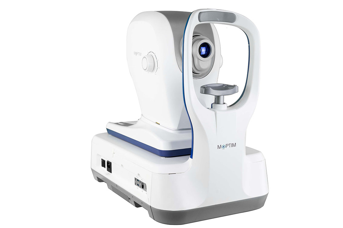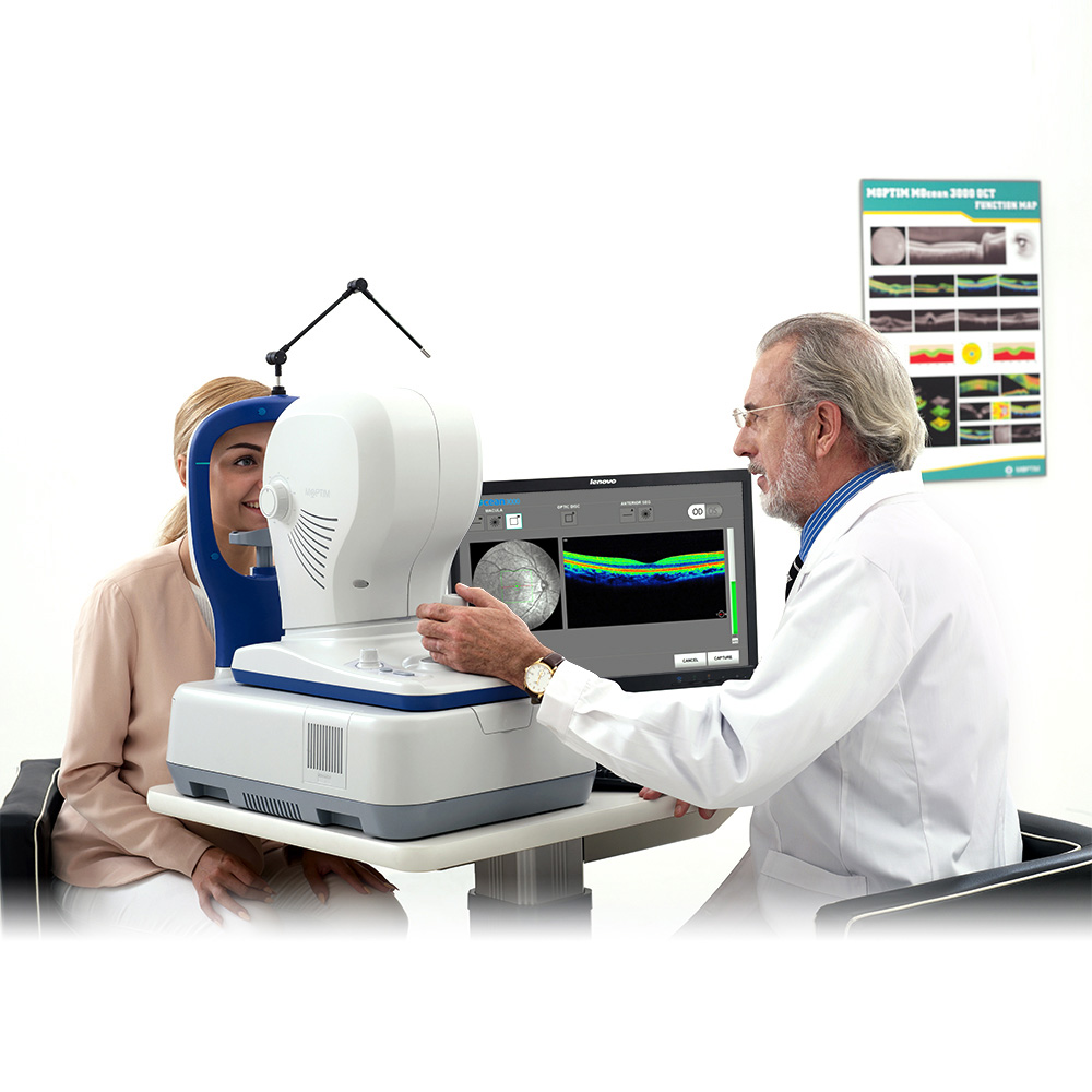
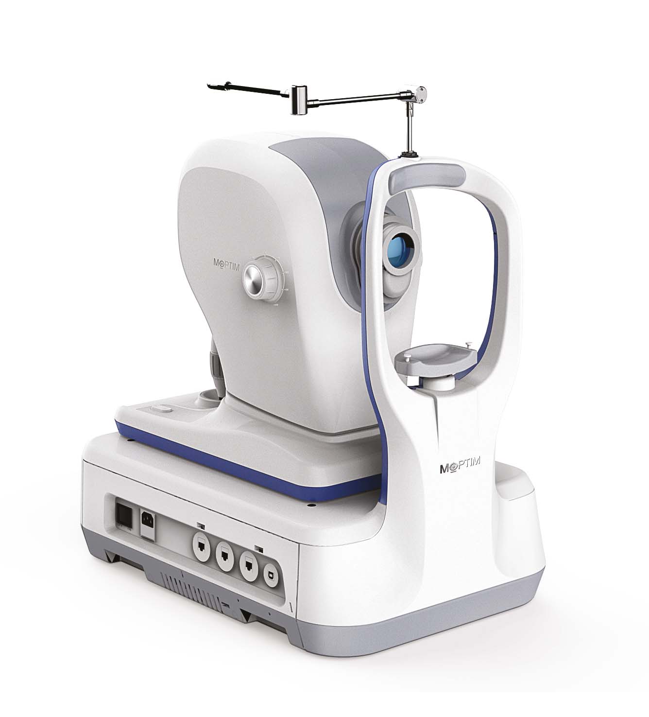
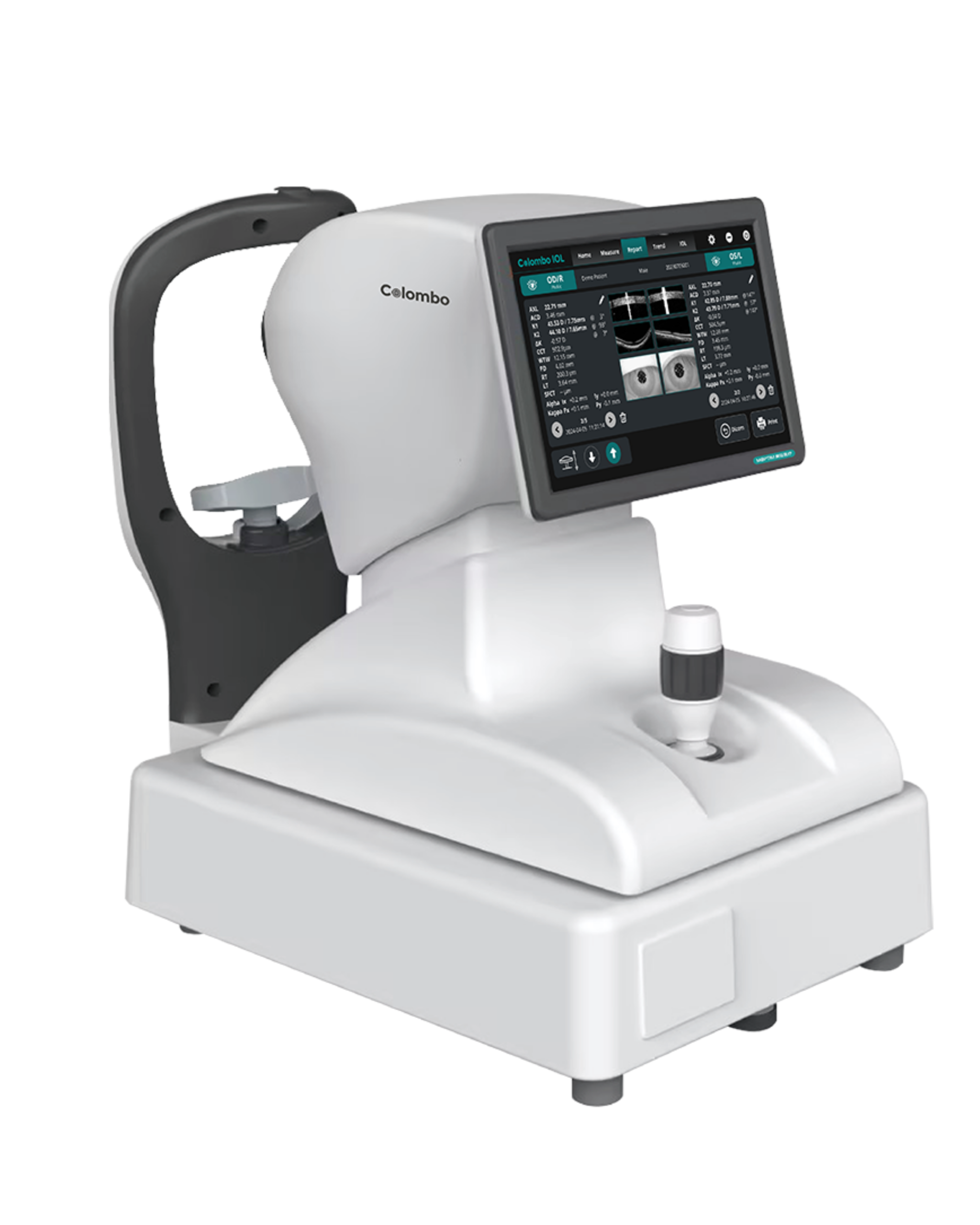
OCT MOCEAN 3000
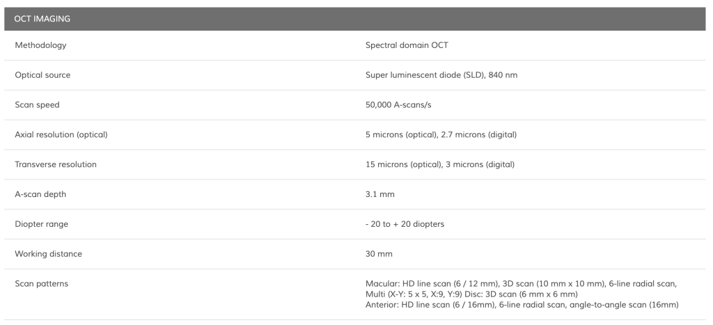
- Scans peed : 50,000 A-scans/s
- Optical source : Super luminescent diode (SLD)
- Axial resolution (optical) : 5 microns (optical), 3.6 microns (digital)
- Transverse resolution : 15 microns (optical), 3 microns (digital)
- A- scan range : 3.1 mm in tissue
- Scan width range : posterior segment 6-12mm , Anterior segment 6-16mm.
Files to download
Image Gallery
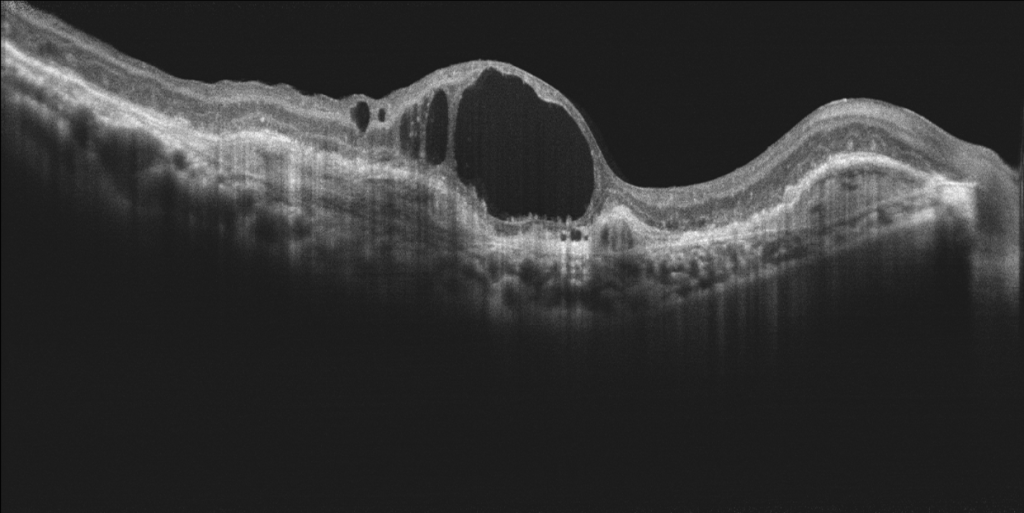
DR
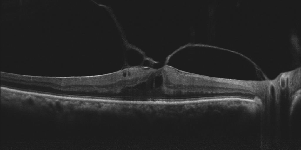
VMT
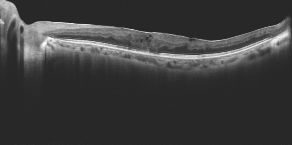
ERM
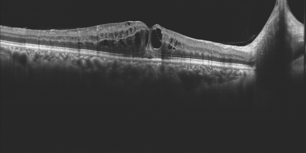
BRVO
Overview
- Up to 10 × 10 mm 3D PanoScan
- 3.1 mm scan depth for enhanced visualization of the vitreous, retina, and choroid
- Real-time 45° SLO retinal imaging with integrated eye tracker
- AI-powered retina analysis
- 16 mm angle-to-angle scan
- Comprehensive analysis of the retina, glaucoma, and cornea
*Please contact us for availability in your country.
Real-time SLO with eye tracking
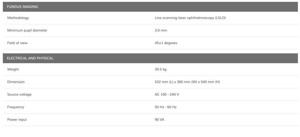
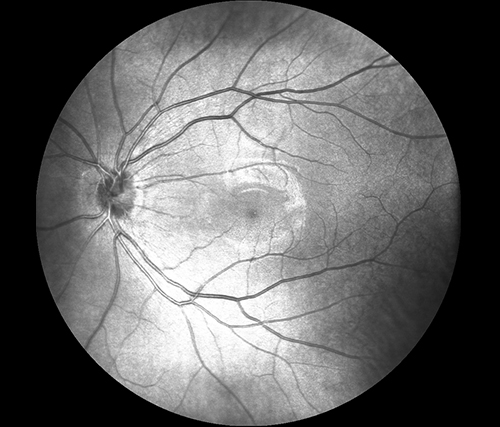
Mocean® 3000 simultaneously acquires OCT images and 45 degrees fundus images based on Scanning Laser Ophthalmoscope (SLO), providing a real-time overview of the retina that allows easy localization of the lesion area before acquisition.
To minimize the artifacts caused by eye drift and micro saccades, Mocean® 3000 uses SLO-based eye tracker, which gives you more confidence in practice.
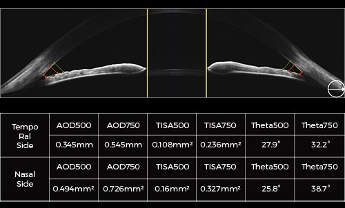
16mm angle-to-angle analysis
16mm angle-to-angle anterior scan with data analysis.
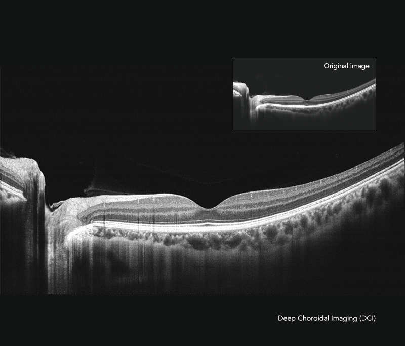
Deep Choroidal Imaging (DCI) mode
Using Deep Choroidal Imaging for detection of choroidal neovascularization.
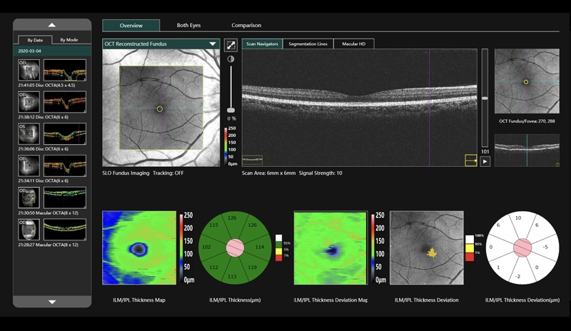
Comprehensive software analysis
The Mocean® 3000 system provides 9 scan patterns to help you improve diagnostic efficiency:
- Retina (HD line, Six-Radial lines, Multi, 3D Cube 10x10mm)
- Glaucoma (Glaucoma Disc for ONH analysis, Glaucoma Macular for GCC analysis)
- Cornea (HD line, Six-Radial lines, Angle-to-Angle)
The software analysis features are always up-to-date and free for upgrade (excluding OCTA module).
SPECIFICATION