OCT
SLO
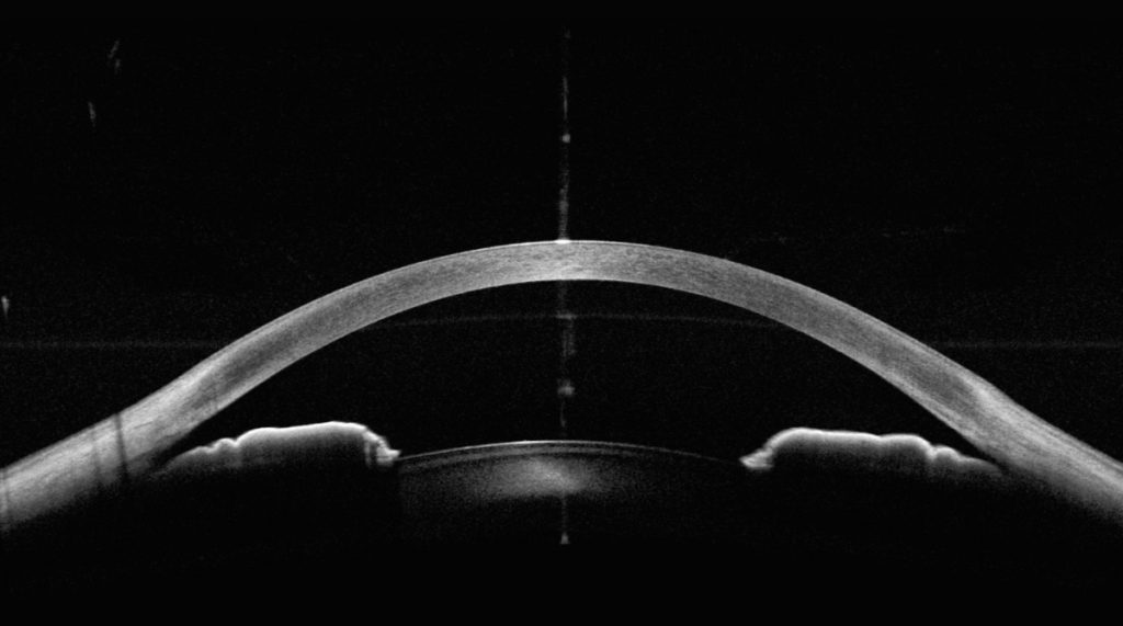
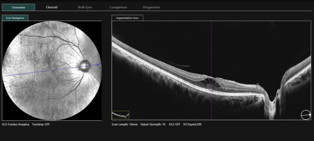
16 mm angle-to-angle scan
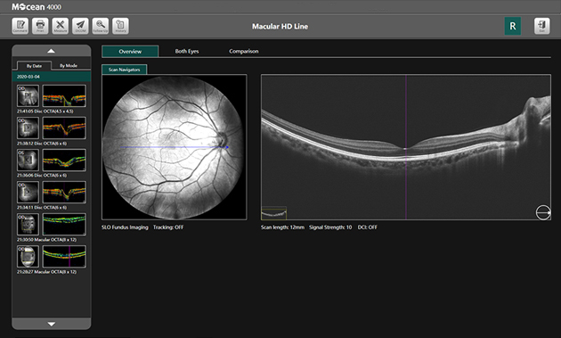
HD scan interface
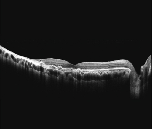
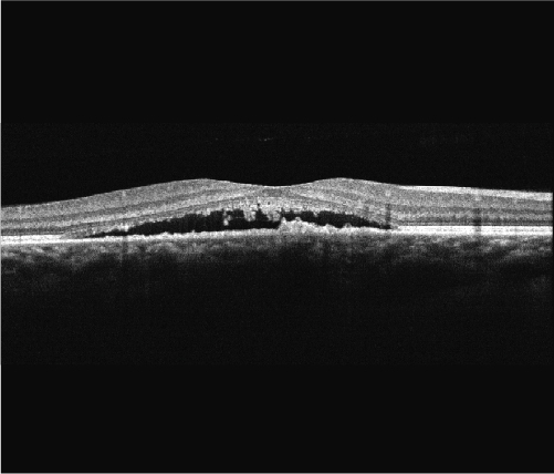
Dry AMD
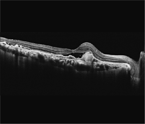
Polypoidal choroidal vasculopathy (PCV)
Central Serous Chorioretinopathy (CSC)
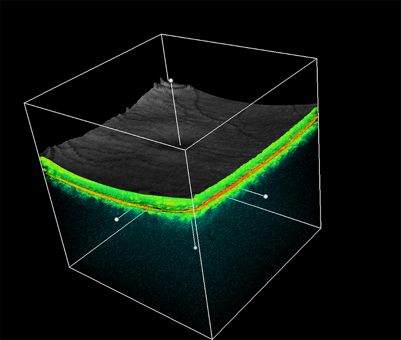
3D view
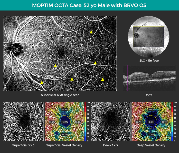
VASCAN OCTA case BRVO
*Availability varies by country. Please contact us for availability in your region
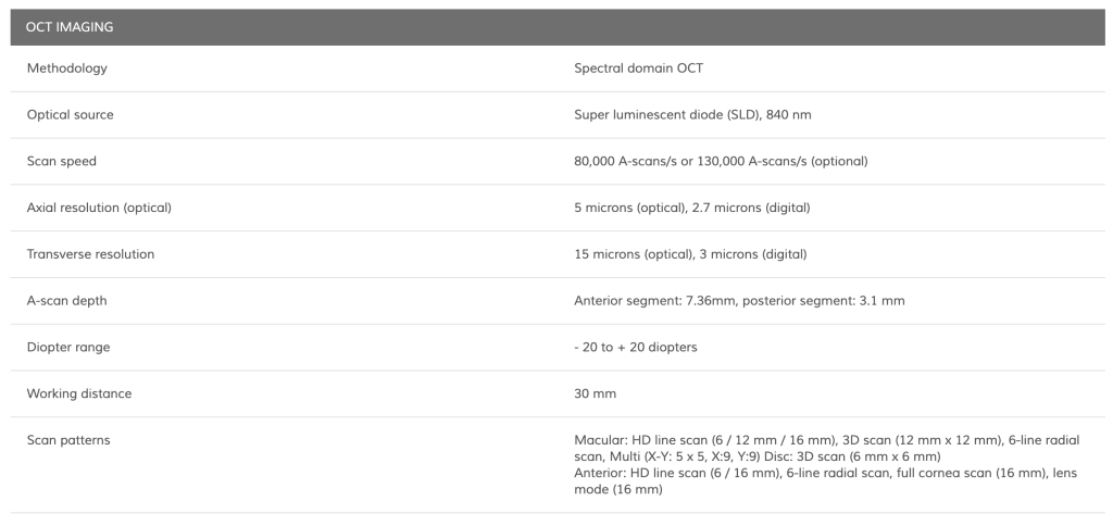
With the capability to perform up to 130,000 A-scans per second, this device ensures fast acquisition of 3D PanoScan images.
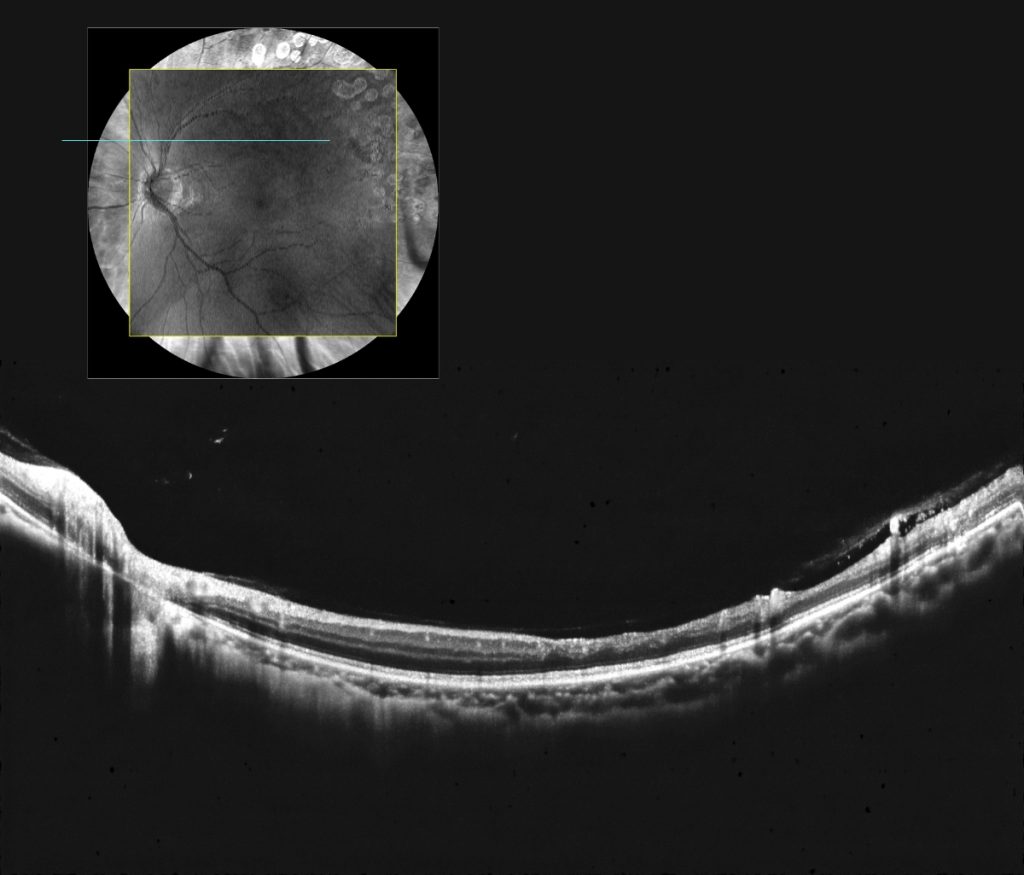
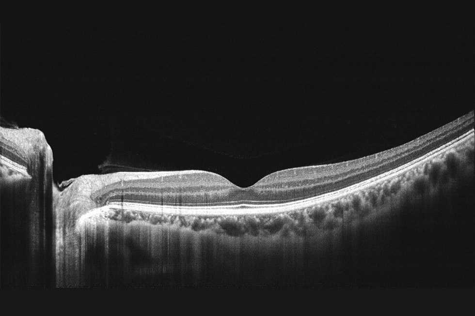
The Mocean 4000’s high definition and widefield 12×12 mm PanoScan feature offers an expansive view in a single scan, revolutionizing the way ophthalmologists assess ocular health. This advanced technology utilizes a unique AI algorithm that significantly enhances image quality, ensuring that no pathological details are overlooked. By delivering a thorough and detailed assessment, the PanoScan feature empowers practitioners with the precision and clarity needed for accurate diagnostics and superior patient care.
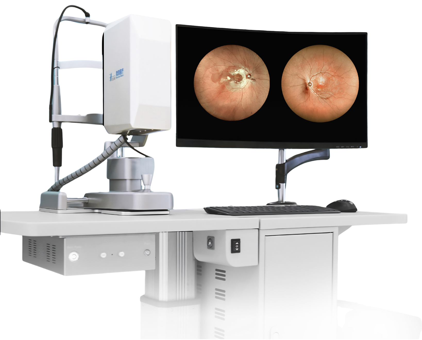
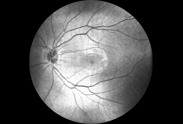
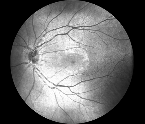
The Mocean® 4000 system stands out with its unique ability to simultaneously capture cross-sectional OCT imaging and 47-degree fundus imaging via Scanning Laser Ophthalmoscope (SLO). This dual capability provides a detailed overview of the retina, making it easier to pinpoint lesion areas prior to acquisition. The system can capture up to 50 SLO fundus images every second, producing high-definition fundus images. To reduce artifacts caused by eye drift and micro saccades, the Mocean® 4000 is equipped with an SLO-based eye tracker that tracks at 100 times per second with an impressive 10-micron accuracy and over a 95% success rate, ensuring greater reliability and confidence in clinical practice.
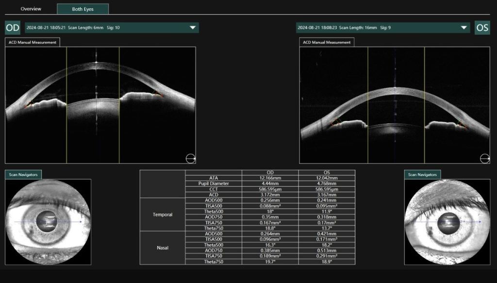
The 16mm full cornea scan feature in Mocean 4000 delivers exceptional and comprehensive imaging of the entire corneal structure. This advanced functionality enables ophthalmologists to capture detailed, high-resolution images, facilitating comprehensive analysis and enhanced diagnostic insights.
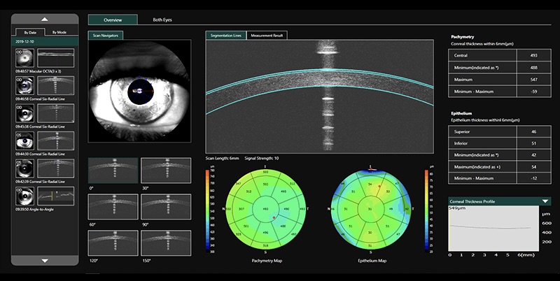
Provides 6mm diameter cornea epithelium thickness map, which is an important part of diagnostics in refractive surgery, with many important clinical applications.
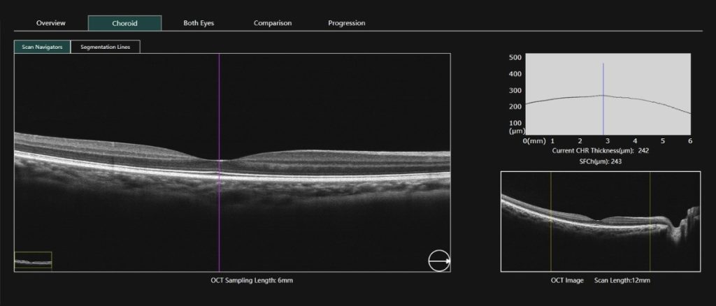
The Mocean 4000 offers automated choroidal thickness analysis along with progression analysis reports, enabling the study of choroidal thickness changes over time.
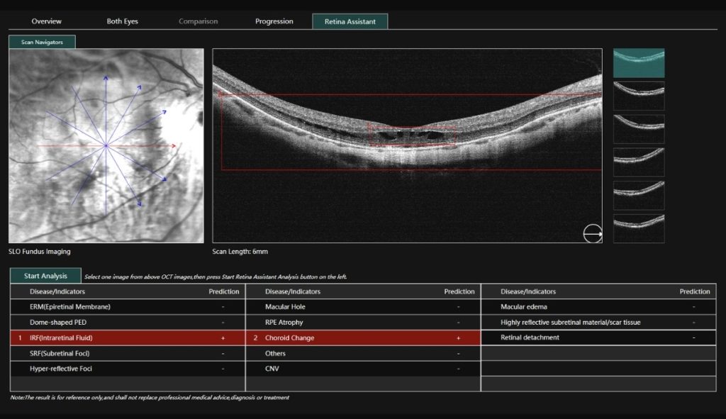
The Mocean 4000 utilizes artificial intelligence for retinal image analysis, assisting doctors in making efficient diagnoses.
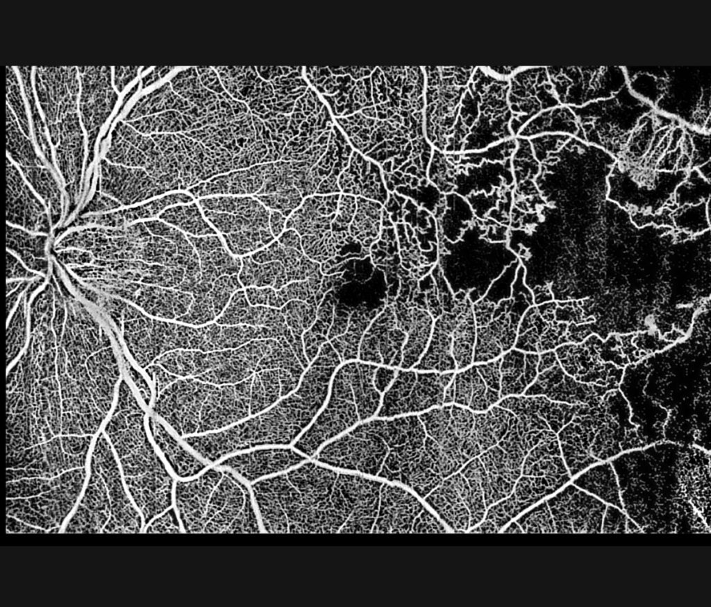
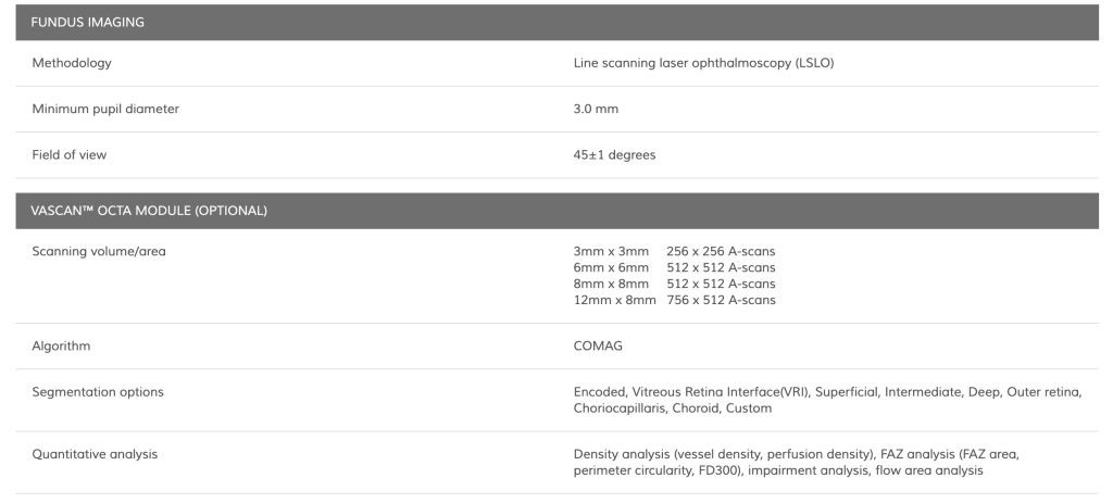
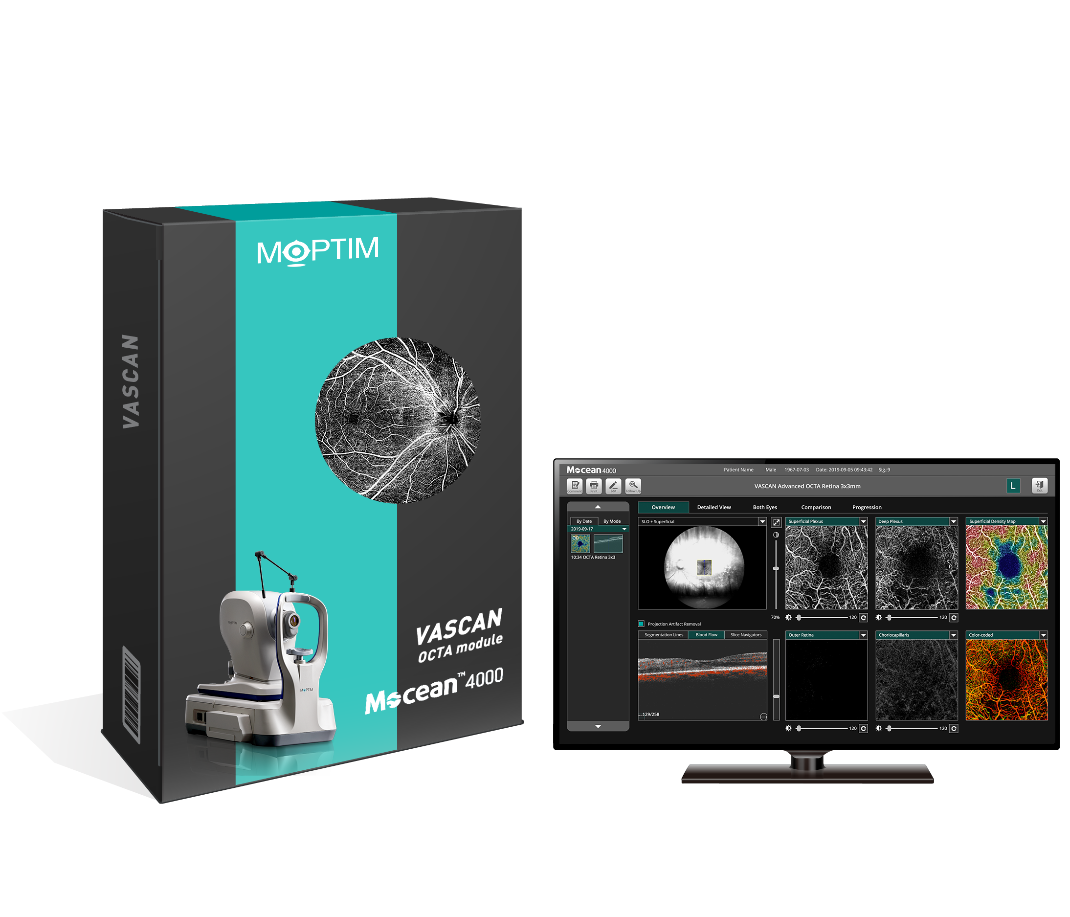
The VASCAN™ OCT Angiography module is an optional software enhancement for the Mocean 4000, offering non-invasive imaging of the eye’s vascular structure without dye injections. It features ultraclear angiographic imaging powered by the COMAG algorithm, delivering widefield OCTA imaging up to 12 mm x 8 mm.
The SLO-based real-time retinal tracking reduces motion artifacts for precise follow-ups, while an advanced projection artifact removal algorithm ensures accurate visualization of retinal vasculature.
The module provides comprehensive quantitative analysis, including density, FAZ, and flow analysis, and supports imaging at various dimensions and slabs, offering detailed views of the retina and optic disc.